
What Is a Stroke?
A stroke happens when blood flow to the brain stops or slows. Because of this, brain cells can die quickly. There are two main types of stroke. One is caused by a blocked blood vessel (ischemic stroke). The other is caused by bleeding in the brain (hemorrhagic stroke). Stroke is a medical emergency. According to the CDC, stroke is a leading cause of death and disability worldwide. Early treatment can save lives and reduce long-term problems.
Why Is Quick Diagnosis Important?
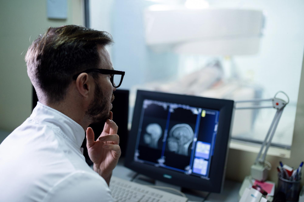
Every minute counts during a stroke. For example, brain cells start to die within minutes. Quick diagnosis helps doctors choose the best treatment. In many cases, fast stroke imaging, like a CT or MRI in stroke, can show what type of stroke is happening. This helps guide the next steps. Without fast imaging, treatment may be delayed. As a result, the risk of long-term damage increases.
What Is a CT Scan?
A CT scan, or computed tomography scan, uses X-rays to make detailed pictures of the brain. During a CT scan for stroke, the machine takes many images from different angles. These images are combined to create a clear picture. CT scans are often the first test done in stroke imaging. They can quickly show if there is bleeding in the brain. This helps doctors know if the stroke is caused by a blocked vessel or by bleeding. Because CT scans are fast, they are used in most emergency rooms.
What Is an MRI?
An MRI, or magnetic resonance imaging, uses strong magnets and radio waves to make pictures of the brain. Unlike CT scans, MRI does not use X-rays. MRI for stroke diagnosis can show more detail than a CT scan. For instance, MRI can spot small or early strokes that a CT scan might miss. It can also show changes in brain tissue. However, MRI scans take longer and may not be available in all hospitals right away.
CT vs. MRI: Key Differences in Stroke Diagnosis
Because of these differences, doctors choose the best test based on each case. Sometimes, both tests are used for a full picture.
How Doctors Choose Between CT and MRI in Stroke Cases
Doctors look at several factors before choosing a scan. For example, if a patient needs fast answers, a CT scan is often used first. If more detail is needed, or if the CT scan is unclear, an MRI may follow. In some stroke imaging centers near you, both scans may be available. Doctors also consider the patient’s health, allergies, and if they have metal in their body. Each case is unique, so the choice depends on what is safest and most helpful for the patient.
What to Expect During a CT or MRI for Stroke
During a CT scan, you will lie on a table that slides into a large, doughnut-shaped machine. The scan usually takes just a few minutes. You may need to hold still, but it does not hurt. Sometimes, a dye is used to help see blood vessels better. For an MRI, you will lie on a table that moves into a tunnel-shaped machine. The scan can take 30 minutes or more. You may hear loud noises, but earplugs are often given. It is important to stay still for clear images. Staff will explain each step before the scan begins.
Safety and Risks of CT and MRI
Both CT and MRI scans are safe for most people. However, there are some risks. CT scans use a small amount of radiation. For most adults, this is not harmful. Pregnant women should tell their doctor before having a CT scan. MRI does not use radiation, but it is not safe for people with some metal implants or devices. In rare cases, the dye used in either scan can cause an allergic reaction. Always tell your doctor about allergies or health problems before the scan.
How Imaging Helps Guide Stroke Treatment
Imaging is key in stroke treatment. For example, CT or MRI in stroke helps doctors see the type, size, and location of the stroke. This information guides treatment choices. If the stroke is caused by a clot, doctors may give medicine to break it up. If there is bleeding, surgery or other treatments may be needed. Imaging also helps doctors check for problems after treatment. Because of this, stroke treatment imaging is an important part of care.
Prevention and Lifestyle Tips After a Stroke Diagnosis
After a stroke, making healthy changes can lower the risk of another stroke. Some helpful tips include:
Many stroke imaging centers near you also offer support and advice for recovery. Making these changes can help protect your brain and overall health.