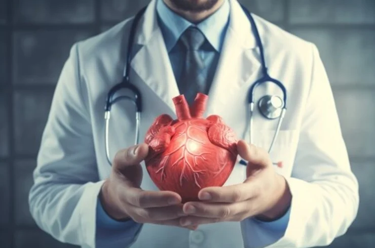
Advanced Visualization for Heart Health: Cardiac Imaging
Cardiac imaging is a state-of-the-art diagnostic technique that enables detailed visualization of the heart’s structure and function. Utilizing technologies such as echocardiography, cardiac MRI, CT coronary angiography, and nuclear scans, this specialized imaging plays a vital role in detecting heart conditions early and accurately. It allows physicians to assess the size, shape, and performance of the heart chambers, evaluate blood flow, identify blocked arteries, and detect congenital or acquired heart diseases. Cardiac imaging is indispensable in diagnosing conditions like coronary artery disease, cardiomyopathy, heart valve disorders, and pericardial diseases. By providing non-invasive, real-time insights into the cardiovascular system, it aids in planning effective treatment strategies and guiding interventions such as angioplasty or bypass surgery.
Precision Diagnosis for Lifesaving Interventions
What sets cardiac imaging apart is its unparalleled precision in uncovering abnormalities that might otherwise go unnoticed until they become life-threatening. With high-resolution imaging, doctors can monitor heart function in patients with diabetes, hypertension, or a history of heart disease. It is also essential in preoperative assessments and post-treatment follow-ups, ensuring the heart is functioning optimally. Safe, painless, and efficient, cardiac imaging is suitable for people of all ages and is often used in both emergency and routine evaluations. By enabling early detection and guiding proactive care, cardiac imaging empowers clinicians to protect heart health and prevent major cardiac events, making it an essential pillar in modern cardiology.